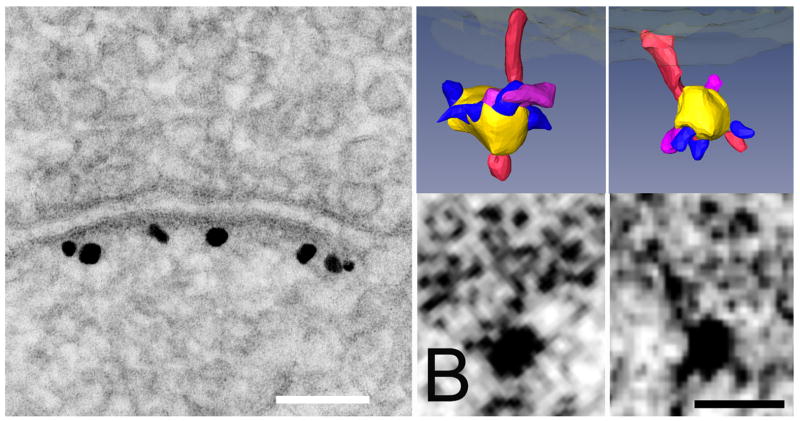
Fig. 1.
(A) Conventional enhanced label for PSD-95 showing high specificity of the polyclonal antibody made to the region of PSD-95 lying between PDZ2 and PDZ3. Scale bar: 100 nm. (B) Parallel specimens of hippocampal culture were silver enhanced for shorter times before preparation for tomography by freeze-substitution, resulting in smaller and less frequent silver grains. Label associated with filaments in single tomographic slices is shown below, and surface renderings above. Vertical filaments are rendered in red, and silver-enhanced gold particles are rendered in yellow. Structures rendered in purple are the correct size to represent the primary antibody contacting the vertical filaments. Smaller structures rendered in blue appear to represent the secondary Fab fragment protruding from the silver encrustation around the original gold particle. Scale bar: 10 nm.

