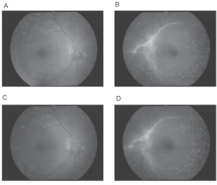
Figure 1.
Fundus photographs before and after pioglitazone treatment. A and B: 4 months before pioglitazone treatment, macula edema is not present in either eye (A shows the right eye, B the left eye). C and D: during pioglitazone, DME is present in both eyes but it is difficult to detect in fundus photographs because the DME is very diffuse (C shows the right eye, D the left eye).
Abbreviations: DME, diabetic macular edema.

