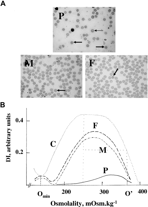
Figure 1.
Blood smears and osmotic gradient ektacytometry. Blood smears (A) and osmotic gradient ektacytometry (B). (A) P indicates proband. Many red blood cells were spherocytes (➡). The remaining showed further changes, as if some fragmentation had occurred. There was a pronounced anisocytosis. A few red cells verged on stomatocytes (→). Erythroblasts were present (17%). F indicates father. Presence of many spherocytes without anisocytosis. M indicates mother. Presence of many spherocytes without anisocytosis. (B) In both parents (M and F), there was an increased osmotic fragility, a reduced maximum deformability index, and a decreased dehydration, a situation typical of HS.11 These features were dramatically enhanced in the proband (P). C indicates control. The maximum deformability index (DImax; normal values, 0.41-0.53 AU) is the maximum value of the deformability index. The “hypo-osmotic point” (Omin; normal values, 143-163 mOsm/L) is the osmolality at which the deformability index reaches a minimum in the hypotonic region; it is the same as the osmolality at which 50% of the erythrocytes hemolyze in a standard osmotic resistance test. This index thus provides a measure of the average surface area–to-volume ratio of erythrocytes. The “hyper-osmotic point” (O′; normal values, 325-375 mOsm/L) is the osmolality in the hypertonic region (right leg of the curve) at which the deformability index reaches half its maximum value. It provides information on the erythrocyte hydration.

