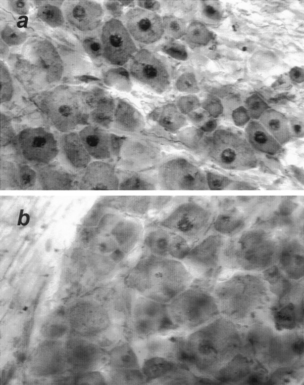
Fig. 3.

Phosphorylated c-Jun is localized to axotomized DRG neurons. Twenty-four hours after proximal nerve transection, axotomized (a) and contralateral untransected (b) DRGs were removed and processed as 35 mm free-floating sections for immunostaining with an antibody specific for serine-63 phosphorylated c-Jun. Intense nuclear staining was restricted to the nuclei of DRG neurons and was strongly evident in axotomized DRGs. Sections shown are representative of immunostaining in DRGs from three separately analyzed animals.
