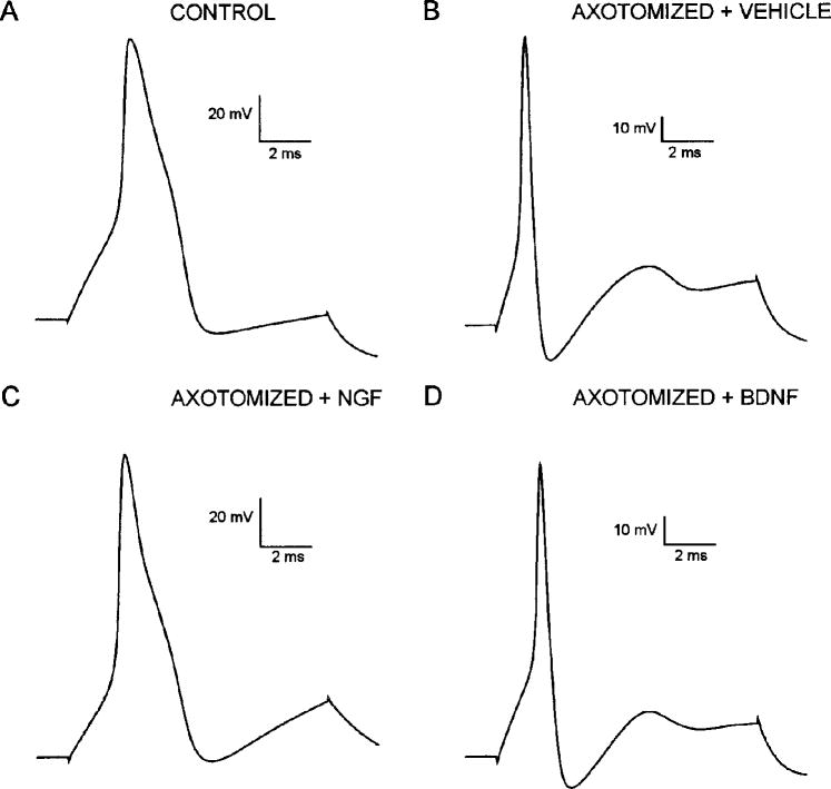
FIG. 3.
AP waveform in control, saline-treated, and neurotrophin-treated neurons after axotomy. APs induced by depolarizing current pulse in control (A) and axotomized neurons after treatment with vehicle solution (B), NGF (C), and BDNF (D). Approximately half of uninjured cutaneous afferent neurons studied had long-duration APs with inflections on downslope (A), whereas 82% of axotomized neurons treated with vehicle solution had short-duration APs lacking this inflection (B). After NGF treatment, 60% of neurons expressed inflected APs (C) and 75% of BDNF-treated neurons lacked inflected APs (D).

