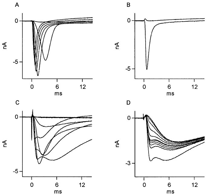
FIG. 7.
Effect of vehicle treatment on Na+ currents in axotomized cutaneous neurons. Na+ currents recorded from 2 DRG neurons whose ligated axons were perfused with vehicle (Ringer) solution. Of 16 neurons studied, Na+ currents in 10 had appearance shown in A. Here, superimposed traces were recorded during test potentials from −40 to +20 mV in steps of 10 mV, after a fixed holding potential of −60 mV. These currents were from a kinetically fast population of Na+ channels completely sensitive to 1 μM TTX (maximal inward current observed at −20 mV: subsequent stimulations have decreasing inward current as reversal potential for Na+ is approached). B: current during test pulses to 0 mV, before, then during, perfusion with TTX. Of 16 vehicle-perfused neurons, 6 had Na+ currents whose complex appearance suggested presence of ≥2 kinetically distinct currents, such as those in C and D. In C, superimposed traces were recorded during test potentials from −50 to +10 mV in steps of 10 mV, after a fixed holding potential of −60 mV. Subsequent, stronger depolarizations revealed rapidly activating and inactivating currents followed by a 2nd, more slowly activating and inactivating component. The 2 components displayed a differential sensitivity to external 1 μM TTX (D). Here, successive depolarizations to +10 mV at 1-s intervals during TTX perfusion show early component of Na+ current to be selectively sensitive (maximum inward current at −20 mV).

