
Fig. 1.
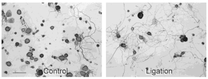
Effects of conditioning lesions on gross appearance of dorsal root ganglion (DRG) cultures. Representative light micrographs of neurofilament stained control (A) and ligation (B) protocol cultures of adult rat DRG neurons after 1 day in vitro. L4 and L5 DRG from the control and conditioning-lesioned side of the animal were dissociated and plated 1 week after subjecting rats to a unilateral midthigh sciatic nerve ligation with the proximal cut end of the nerve pulled through a silicon-plugged cuff to prevent regeneration through the denervated nerve segment. Note the enhanced neurite outgrowth in the culture exposed to a previous axotomy in vivo. Scale bar = 100 μm.