
Fig. 1.
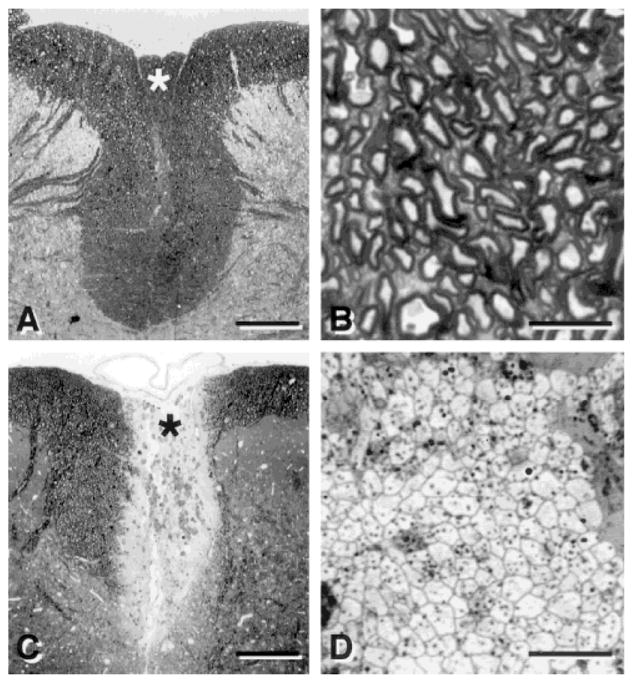
Light micrographs of transverse sections of the dorsal spinal cord showing the dorsal funiculus in normal (A) and demyelinated (C) rats. Examination at higher magnification shows normal (B) and demyelinated (D) axons in the dorsal columns. B and D were prepared from the area around the asterisk in A and C, respectively. Scale bar = 250 μm in A,C; 10 μm in B,D.