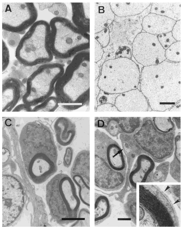
Fig. 4.
Electron photomicrographs showing normal (A) and demyelinated (B) axons in the dorsal columns. All demyelinated spinal cords that received human OEC injections showed clear evidence of remyelination (C) of the demyelinated axons. Some human OECs ensheathed two or more axons (D; arrow). Examination at higher magnification showed the presence of a basal lamina surrounding the fibers characteristic of peripheral myelin (inset, arrowheads). Scale bars = 1 μm in A, 3 μm in B, 2 μm in C, 1.5 μm in D.

