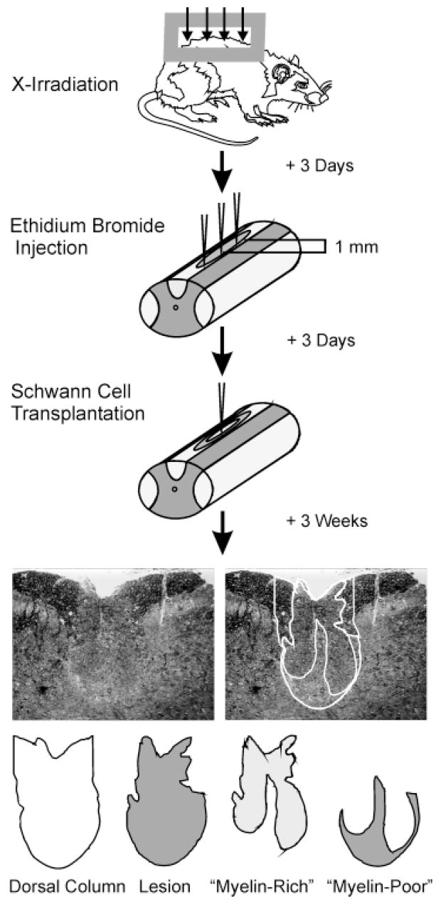
Fig. 1.
Diagram illustrating the basic protocol used in this study. Demyelinating lesions were created in the dorsal columns of adult rats using X-irradiation and ethidium bromide injections. Cells were transplanted into the centers of lesions, and rats were sacrificed 3 weeks after transplantation. Spinal cords were processed for histological analysis, and cross-sectional areas of the dorsal funiculus, the lesion, and “myelin-rich” and “myelin-poor” areas within the lesion were measured and densities of myelinated axons were counted within each region at 0.25-mm intervals along the spinal cord. These values were used to calculate the total amount of axon length remyelinated and to estimate the percentage of myelin restoration.

