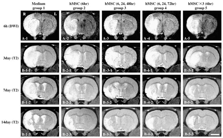
Fig. 1.
Evaluation of the ischemic lesion volume with Diffusion Weighted Images (DWI) and T2Weighted Images (T2WI). hMSCs were intravenously injected at various times and doses after the initial MRI scan (6 h after MCAO). DWI obtained at 6 h after MCAO in group 1 (A1), group 2 (A2), group 3 (A3), group 4 (A4), and group 5 (A5). T2WI obtained at 3, 7 and 14 days after MCAO in group 1 (B1-1 to 3), group 2 (B2-1 to 3), group 3 (B3-1 to 3), group 4 (B4-1 to 3), and group 5 (B5-1 to 3). Scale bar=5 mm.

