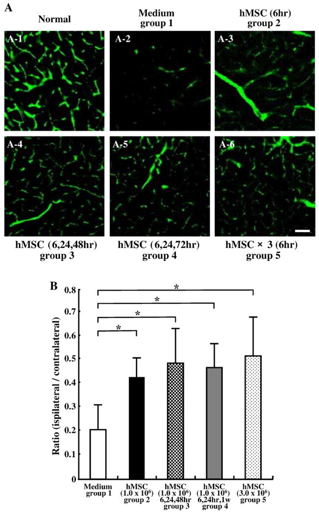
Fig. 5.
Fourteen days after MCAO, angiogenesis in boundary zone was analyzed using a three-dimensional analysis system. Panel A-1 shows the three-dimensional capillary image with systemically perfused FITC-dextran in the normal rat brain. The capillary vascular volume was expressed as a ratio by dividing that obtained from the ischemic hemisphere by that of the contralateral control hemisphere. At 14 days after MCAO, the ratio (ipsilateral/contralateral) was increased in group 2 (A-3), group 3 (A-4), group 4 (A-5), and group 5 (A-6) compared to group 1 (A-2). These results were summarized in panel B. Scale bar=150 μm. *P<0.05.

