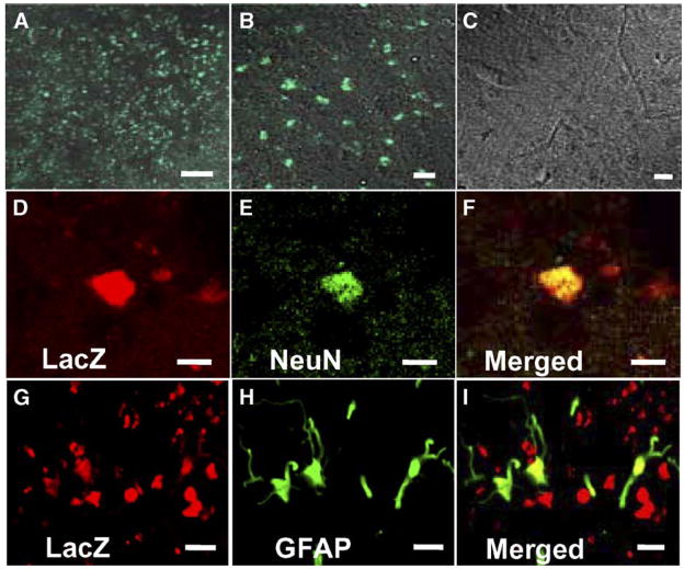
Fig. 4.
Intravenously-administrated hTERT-MSCs accumulated in and around the ischemic lesions. Lower (A) and higher micrograph (B) demonstrating a large number of eGFP-positive cells in and around the lesion, but not in the non-treated rats (C). Confocal images show the differentiation of the transplanted hTERT-MSCs (LacZ in red; D, G) into neurons (NeuN in green; E) or astrocytes (GFAP in green; H). Panels F and I confirm the co-labeling of LacZ/NeuN or LacZ/GFAP in the cells, respectively. Scale bar = 250 μm (A), 10 mu;m (B, C), 5 mu;m (D–I).

