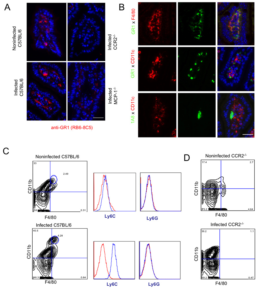
Figure 3. Recruitment of Gr1+ (Ly6C+) monocytes to the lamina propria in the ileum following T. gondii infection depends on CCR2 and MCP-1.
(A) Recruitment of Gr1+ cells in the ileum of orally infected C57BL/6 mice. Gr1+ cells increased within the lamina propria and were found beneath the basement membrane, surrounding the villus core within the ileum. Gr1+ cells were also seen in noninfected wild type (C57BL/6) but not in noninfected (data not shown) or infected CCR2−/− and MCP-1−/− mice. Frozen sections of orally infected mice were examined 9 days post-infection with anti-Gr1 (RB6-8C5) followed by Alexa-594 conjugated goat anti-rat IgG. Scale bar = 25 µm.
(B) Characterization of Gr1+ cells in the lamina propria of orally infected mice. Gr1+ cells stained with F4/80 but not CD11c or the neutrophil marker 1A8. Frozen sections of ileum from orally infected mice were examined after 9 days post-infection. Top panel: anti-Gr1 (RB6-8C5) directly conjugated to Alexa 488 and anti-F4/80 directly conjugated to Alexa 594; middle panel: anti-Gr1 (RB6-8C5) and anti-CD11c (N418) followed by Alexa-conjugated secondary antibodies; Bottom panel: anti-Ly6G (1A8) and anti-CD11c (N418), followed by Alexa-conjugated secondary antibodies Scale bar =25 µm.
(C) Oral infection with T. gondii induced recruitment of Gr1+ (Ly6C+) monocytes to the ileum. Leukocytes were isolated from the ileum of mice on day 9 after oral infection, stained for CD11b (mAb M1/70 conjugated to PE or FITC) and F4/80 (mAb A31 conjugated to APC), and quantified by FACS. Double positive macrophages (CD11b+ F4/80+) were gated based on isotype controls and circled populations were analyzed after staining with anti-Ly6C (mAb AL-21 conjugated to FITC) or Ly6G (mAb 1A8 conjugated to PE). Resident macrophages in non-infected mice (top) were negative for both Ly6C and Ly6G. In contrast, doubly positive cells from infected mice are strongly positive for Ly6C, but did not stain for Ly6G (bottom). Results shown are representative of three or more experiments; cells were pooled from three mice in each group.
(D) Analysis of peritoneal cells in noninfected (top) and infected (bottom) CCR2−/− mice. While neutrophils (CD11b+, F4/80−) increased dramatically in infected mice, the population of doubly positive CD11b+ F4/80+ cells was absent.

