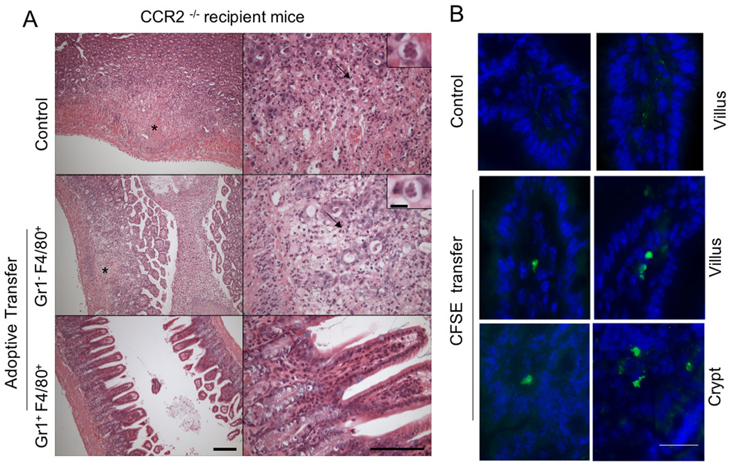
Figure 6. Adoptively transferred Gr1+ (Ly6C+) monocytes home to the intestine and prevent extensive pathology in CCR2−/− mice.
(A) Sorted cells were adoptively transferred to infected CCR2−/− mice on day 3 post-infection and the ilea were examined at day 9 by H&E staining of paraffin sections. Typical lesions (asterisk) were observed in the ileum of infected CCR2−/− mice that did not receive cells (Control) or received F4/80+ singly positive cells. CCR2−/− mice that received Gr1+ (Ly6C+) F4/80+ cells show well-preserved architecture in the ileum, similar to wild type mice. Representative of two similar experiments containing 2 animals per group. Scale bar = 200 µm (15 µm inset). Right panels are enlarged views.
(B) Adoptively transferred Gr1+ (Ly6C+) monocytes home to the ileum of infected mice. Sorted, CFSE labeled Gr1+ (Ly6C+) F4/80+ monocytes i.v. inoculated into C57BL/6 mice on day 3 after oral infection. One day following adoptive transfer (day 4 after oral infection), ilea were collected and frozen sections were examined by fluorescent microscopy. Recruitment of CFSE labeled Gr1+ (Ly6C+) monocytes was observed in the lamina propria region in the villi and in the crypt region, which contained lymphocytic aggregations. Control represents C57BL/6 mouse that was infected but did not receive CFSE labeled cells. Scale bar = 20 µm.

