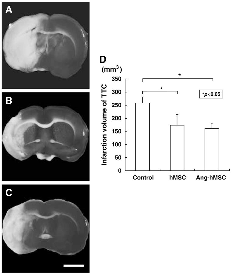
Figure 3.
TTC Brain sections slices stained with TTC to visualize the ischemic lesions 7 days after MCAO. 2,3,5-Triphenyl tetrazolium chloride-stained brain slices from (A) mediuminjected MCAO model rats, (B) following hMSC-treated, and (C) Ang-hMSC-treated groups. (D) Seven days after MCAO, there was a reduction in the lesion volume assayed using TTC staining for both hMSC and Ang-hMSC groups. Bar=3 mm.

