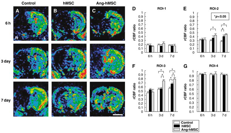
Figure 6.
Evaluation of hemodynamic state (rCBF maps) with PWIs. Human mesenchymal stem cells or Ang-hMSCs were intravenously injected immediately after the initial MRI scanning (6 h after MCAO). Images obtained 6 h, 3, and 7 days MCAO in (A) medium-injected, (B) hMSC-treated, and (C) Ang-hMSC-treated group. (D–G) Summary of rCBF evaluated with PWI in each groups. (D) ROI-1, (E) ROI-2, (F) ROI-3, and (G) ROI-4. Regional cerebral blood flow ratio (ischemic lesion/contralateral lesion) at 6 h, 3, and 7 days after MCAO are summarized in D–G. Bar=3 mm, *P<0.05.

