
Fig. 2. Cell cycle arrest and cyclin D1 degradation induced by NiCl2, but not NiS, in lung A549 cells.
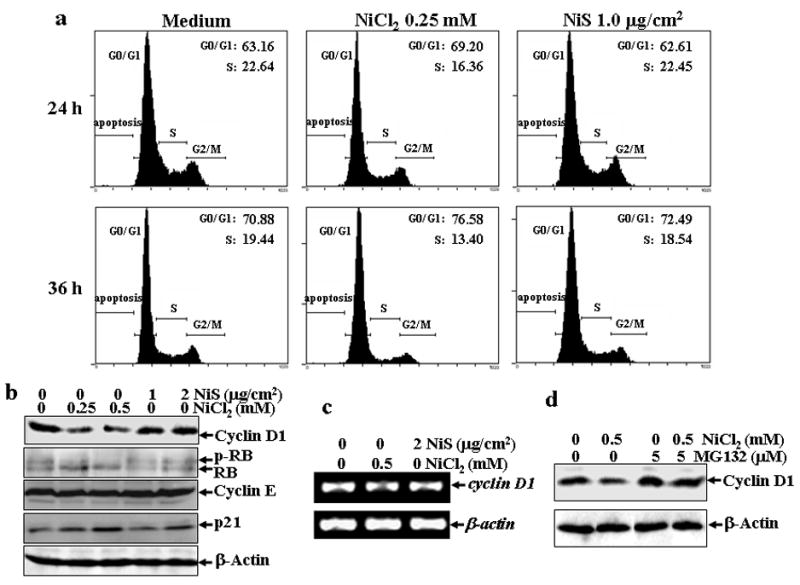
(a), A549 cells were seeded into each well of 6-well plates and cultured in Ham’s F-12K containing 10% FBS. After the cell density reached 70–80%, the cells were exposed to 0.25 mM NiCl2 or 1 μg/cm2 NiS for 24 or 36 hrs, and then were fixed and stained with propidium iodide as described in Materials and Methods. Cell cycle distribution was determined by flow cytometry. The data presented in this figure indicating the percentage of G0/G1 and S phases was one representative of three independent experiments. (b and d), The cells were treated with various concentrations of nickel compounds as indicated for 24 h (b), or 0.5 mM of NiCl2 in the presence or absence of MG132 for 12 h (d), and then extracted with SDS-sample buffer and Western blot was carried out. β-actin was used as a control for protein loading control. (c), The cells were exposed to 0.5 mM NiCl2 or 2 μg/cm2 NiS for 12 h, and then extracted with Trizol reagent for the total RNA isolation. Cyclin D1 was amplified with its specific primers by RT-PCR for 25 cycles. β-actin was used as an internal control.