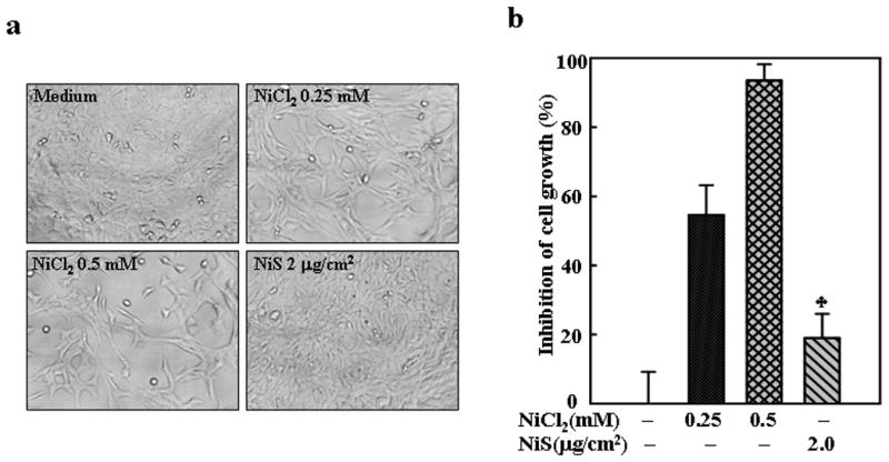
Fig. 3. Inhibition of MEFs growth by NiCl2.

1 × 10³ of MEFs were seeded into each well of 96-well plates, cultured in 10% FBS DMEM overnight, and then exposed to NiCl2 or NiS for 72 h. The cells were photographed under microscopy (a), and then extracted by lysis buffer for growth measurement using CellTiter-Glo® Luminescent Cell Viability Assay kit with a luminometer (b). The results are expressed as inhibition of cell growth which was calculated as described in Material and Methods. The symbol (♣) indicates a significant different from NiCl2 group (p< 0.05).
