
Fig. 6. IKKα is a mediator for cyclin D1 degradation and cell cycle arrest induced by NiCl2.
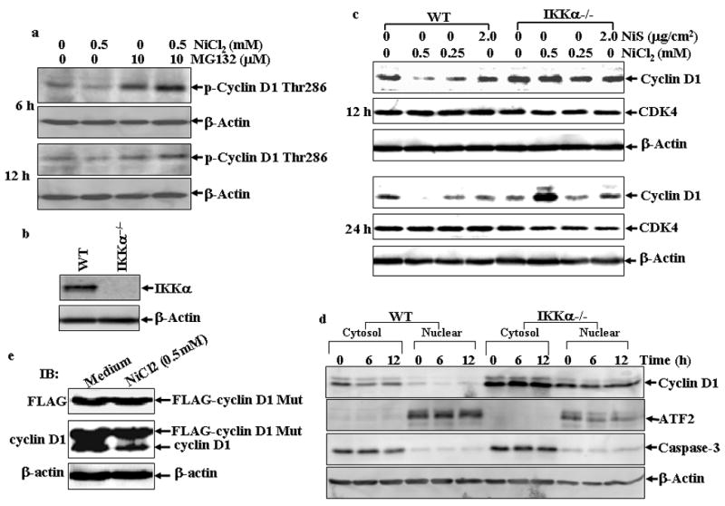
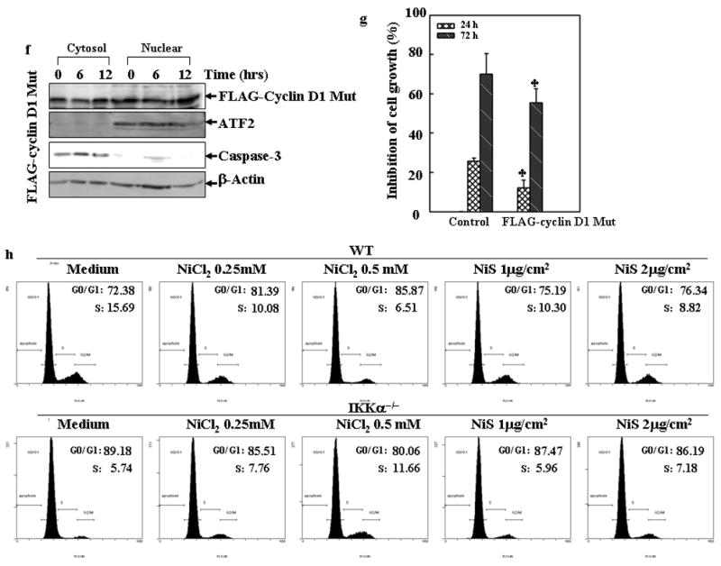
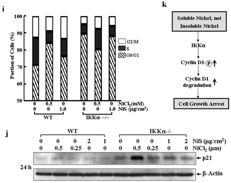
(a), A549 cells were treated with 0.5 mM of NiCl2 in the presence or absence of MG132 for 6 h and 12 h, and then extracted with SDS-sample buffer and Western blot was carried out. (b), IKKα−/− MEFs were identified by Western blot. (c), WT MEFs and IKKα−/− MEFs were seeded into each well of 6-well plates and cultured in DMEM containing 10% FBS at 37°C overnight. The cells were exposed to various concentrations of nickel compounds for 12 or 24 hrs, and then extracted with SDS-sample buffer. The cyclin D1 protein levels in the extracts were analyzed by Western blot. CDK4 and β-Actin were used as controls. (d), WT MEFs and IKKα−/− MEFs were seeded into 10-cm dishes and exposed to various concentrations of nickel compounds for 6 or 12 h, and then the nuclear and cytosol parts were extracted as described in “Materials and Methods”. The cyclin D1 protein levels in the extracts were analyzed by Western blot. ATF2 and caspase-3 were used as nuclear and cytosol marks individually. (e–g), 293T cells were transiently transfected with FLAG-cyclin D1 T286A expression vector as described in “Material and Methods”. 24 hrs after the transfection, the cells were treated with 0.5 mM NiCl2 for 12 hrs and then the FLAG-cyclin D1 T286A mutant and endogenous cyclin D1 protein in the extracts were analyzed by Western blot assay with antibodies against FLAG and cyclin D1 (e). Then the subcellular location of the FLAG-cyclin D1 T286 mutant was detected (f). The growth inhibitory effect of NiCl2, was compared between the vector control and FLAG-cyclin D1 T286 mutant cells. Each bar indicates the mean and standard deviation of triplicate assay wells. The symbol (♣) indicates a significant compared with vector control cells (p< 0.05) (g). (h–j), WT MEFs and IKKα−/− MEFs were seeded into each well of a 6-well plates and cultured in DMEM containing 10% FBS at 37°C overnight. The cells were exposed to various concentrations of nickel compounds as indicated for 24 hrs, and then fixed and stained with propidium iodide. The cell cycle distribution was determined by flow cytometry (h & i). The data presented in this figure indicating the percentage of G0/G1 and S phases was one representative of three independent experiments, or extracted with SDS sample buffer, and the cell extracts were analyzed by Western blot for p21 protein expression (j). β-Actin was used as protein loading control. (k), The overall scheme of the inhibitory effect on cell growth by soluble, but not insoluble nickel compounds.