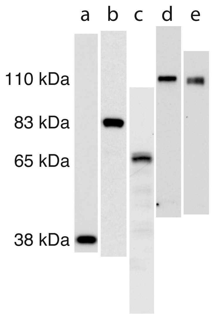
Figure 1. Antibodies used for immunohistochemistry recognize a single band in juvenile superior colliculus.
Western blots from 4–14% poly-acrylamide gels after running denatured, whole lysate of the superficial visual layers of superior colliculus of P14 rats. The scans of lanes have been approximately aligned to show the relative kDa of bands. The lanes are as follows: (a) synaptophysin, (b) synapsin-1, (c) GAD 65/67, (d) GluR1, (e) GluR2. The GAD 65/67 antibody recognizes a doublet as expected, while the others recognize a single band as can be seen in the figure that includes the entire length of each lane.

