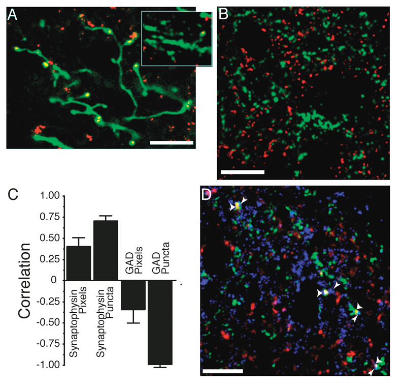
Figure 3. Immunohistochemistry for synapse associated proteins can be used to count retinal ganglion cell axon synapses.
A. The pattern of synaptophysin staining within an axon is consistent with its identity as a synapse. The image is a collapsed confocal z-series from a 10 µm depth of tissue. Synaptophysin puncta occur in expansions of the axon either at terminal points or en passant. This image was taken within the stratum opticum of the ipsilateral colliculus where the axonal caliber is larger and synaptic density is lower. This allows for reconstruction over greater depths to visualize the entire arbor without interference from other axons. Other images presented in this paper are single confocal sections, which is required because of the density of contralateral axons in the SGS (for example, Fig. 2). A single confocal section from the contralateral retinal projection zone that contains a portion of arbor is shown in the inset. The localization of synaptophysin is similar to that seen in the ipsilateral projection, where the exact localization can be better ascertained. B. Sample confocal micrograph showing double labeling of retinal axons (green) and GAD 65/67 (red). Unlike the synaptophysin double labeling (Fig. 2B), there was no overlap between the two labels here. Even though our antibody recognizes GAD 65 and GAD 67, the majority of staining was restricted to processes with only light staining of cell bodies, indicating that GAD 65 was primarily localized during these overlap experiments. C. Correlation coefficients of overlap between ipsi axons and the immunostain for synaptophysin or GAD 65/67. The GAD stain is used as a control because it should never co-localize with retinal axons. Positive correlation indicates that the overlap of axon and antigen is greater than would be expected by chance (see Methods). This correlation coefficient was determined for the total number of overlapped “Pixels”, as well as for the number of pixel accumulations that exceeded 0.16 µm2 (“Puncta”), our minimal criterion for a synapse. Synaptophysin antigens show more overlap, and GAD-65/67 localization shows less overlap, than would be expected by chance, especially when the minimal size criterion is imposed. Data for each antigen were acquired from 12 sections from two animals. D. Overlapped synaptophysin puncta are adjacent to post-synaptic markers. A confocal micrograph of tissue triple labeled for the ipsilateral RGC axons (green), synaptophysin (red – overlap is yellow), and also for the post-synaptic glutamate receptor GluR1 (blue). Presumptive synapses are marked with wide arrowheads. All identified presumptive synapses are adjacent to at least one GluR1 puncta that does not overlap with the retinal axon (thin arrowheads). Scale bars 10 µm.

