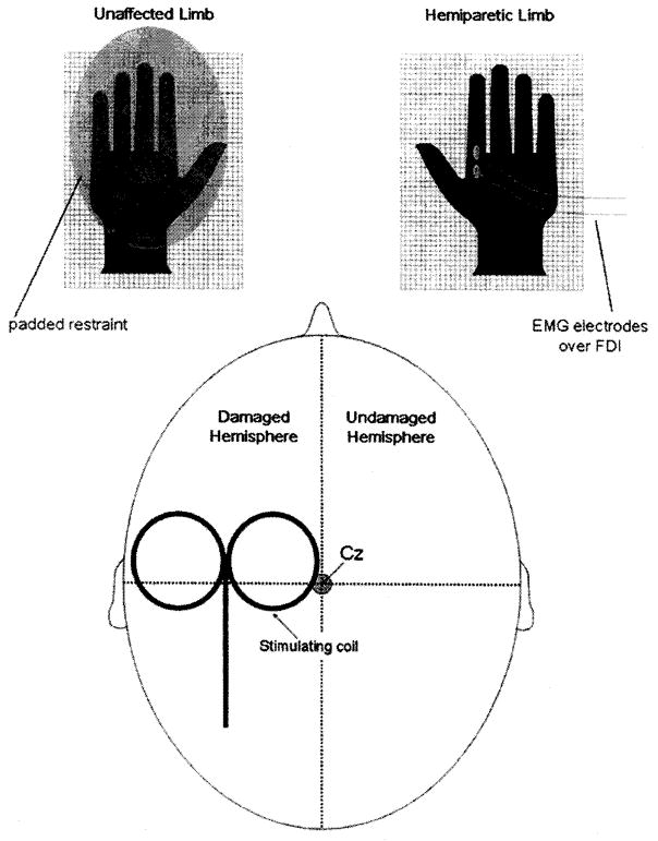
FIGURE 1.
Schematic of rTMS set-up. The magnetic stimulating coil was positioned over the motor cortex of the damaged hemisphere and 2000 stimuli were administered as 50 trains of 40 stimuli, stimulus rate of 20 Hz, stimulus train duration of 2 secs, with an intertrain interval of 28 secs. Passive bipolar electromyographic surface electrodes were applied over the FDI for the purpose of monitoring muscle activation during and between stimulations.

