Figure 4.
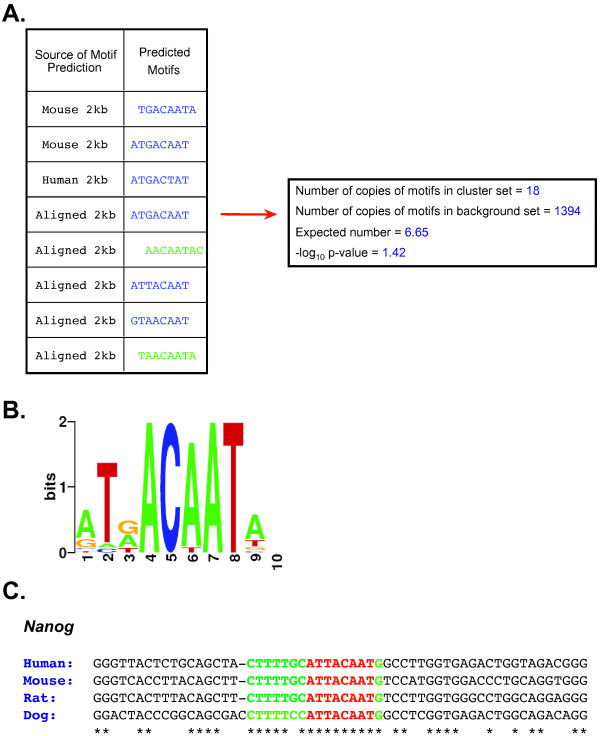
CompMoby identification of the Oct4 and Sox2 binding site upstream of Nanog in mouse embryonic stem cells. (A) The table shows the sequence source of the motifs in the motif cluster. CompMoby predicted the reverse complement of the motifs in green. The right box shows the number of occurrences of the motif cluster in the 2kb upstream sequences of the co-regulated mouse pluripotent gene set compared to a background set of ~8500 other mouse genes. The expected number is calculated from the number of occurrences in the background set, assuming a random distribution. The -log10 p-value is Bonferroni corrected by the total number of motif clusters. (B) WebLogo (v. 2.8) [47] representation of the motif cluster. (C) Four species alignment [27] of upstream region of Nanog containing the Oct4 and Sox2 binding sites. Red represents the motif predicted by CompMoby. Green represents the flanking region extended from the predicted motif based on conservation of the nucleotide position in three out of four species. Asterisk (*) represents a fully conserved position.

