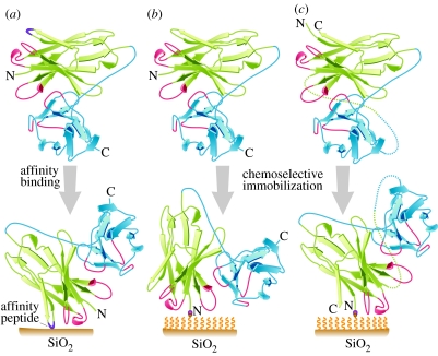
Figure 3.
Ribbon diagrams of scFvs (VH–VL configuration) to be deposited on a SiO2 surface. VH is in green, VL is in blue. Complementarity determining regions (CDRs) of VH and VL are in red. SiO2 surface is represented by a brown bar. A polymeric SAM on a SiO2 surface is represented by wavy yellow lines. N- and C-ends of scFvs are indicated. Chemoselective ligation between N-ends of scFvs and SAM is indicated. (a) Affinity peptide-scFv: SiO2 affinity peptide selected from a display library (Eteshola et al. 2005) is inserted into the scFv antibody fragment (purple line). The affinity peptide binds the SiO2 surface, effectively orienting the scFv and determining the proximity of the CDRs to the SiO2 surface. (b) Parent scFv: chemoselective conjugation of a modified scFv (with an N-terminal aldehyde; Lee et al. 2004) to oxyamine residues on the SAM. Note position of VH CDRs. (c) CP scFv: chemoselective conjugation of a circularly permuted (CP; Eteshola et al. 2006), but otherwise comparable, scFv. In (b) and (c), note that chemoselective conjugation produces a consistent orientation of scFvs, and that, relative to the parent scFv, CP alters the proximity of the CDRs to the SiO2 surface (accepted JRSI-2007-1033).

