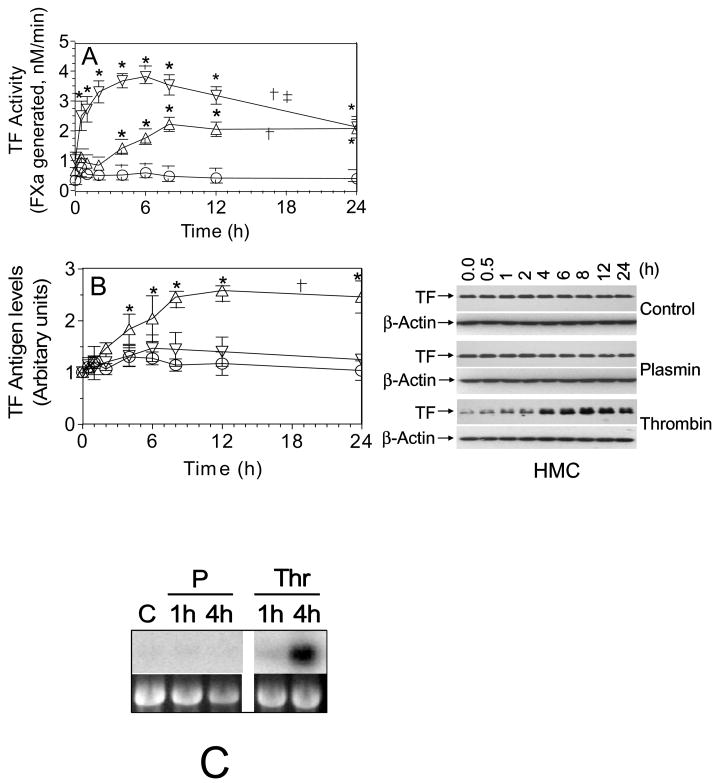
Fig. 1.
Upregulation of TF coagulant activity on human mesothelial cells exposed to plasmin or thrombin. Quiescent HMC monolayers were exposed to control vehicle (○), plasmin, 50 nM (▽) or thrombin, 5 nM (△) for varying time periods. At the end of the treatment period, plasmin or thrombin was removed and the cells were washed twice in buffer A and the cell surface TF activity (A) and TF antigen (B) were measured. Values are shown as mean ± SEM (n=3 to 6 experiments). The right panel shown in Fig 1B was a representative TF immunoblot. The data were analyzed by repeated measures one-way ANOVA followed by Dunnet’s post-hoc test. In panel A, † denotes TF activity over time in plasmin- and thrombin-treated cells significantly differs from that of control vehicle-treated cells (p < 0.01). ‡ denotes TF activity overtime in plasmin-treated cells significantly differs from that of thrombin-treated cells (p < 0.001). In panel B, † denotes TF antigen over time in thrombin-treated cells significantly differs from that of control vehicle- or plasmin-treated cells (p < 0.01). TF antigen over time in plasmin-treated cells does not significantly differ from that of control vehicle- treated cells. * represents the value significantly differs from the value noted at zero time (p < 0.05). (C) Effect of plasmin and thrombin on HMC TF mRNA expression. Quiescent HMC were treated with serum free medium alone for 4 h (C), plasmin (P), (50 nM) or thrombin (Thr), (5 nM) in serum free medium for 1 h or 4 h. Total RNA was isolated from the cells, 10 μg of each RNA sample was used for Northern blot analysis, and the blot was hybridized with a radiolabeled TF cDNA probe and exposed to X-ray film. Ethidium bromide staining of 18S ribosomal RNA of the same samples was shown for RNA loading control.

