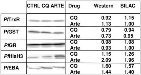
Figure 4. Validation of SILAC data by immunoblotting.
The following polyclonal antibodies were used: rabbit-anti-PfTrxR (thioredoxin reductase), rabbit-anti-PfGST (glutathione S-transferase), rabbit-anti-PfGR (glutathione reductase) (available in the Becker Lab), rabbit-anti-Histone H3 (Abcam, Cambridge, UK), rabbit-anti-PfEBA (Erythrocyte Binding Antigen) (MRA-2 by MR4) at appropriate dilutions 1∶1,000–5,000 and peroxidase-conjugated anti-rabbit antibody (Dianova, Hamburg, Germany, anti-IgG, 1∶50,000–100,000). Visualization was performed by enhanced chemiluminescence (ECL, SuperSignal West Femto Maximum Sensitivity Substrate, Pierce Biotechnology, Rockford, IL, USA). Densitometric quantification was performed using the Quantity One software (BioRad) by quantitating band volumina (trace intensity×mm2) of each sample and comparing treated and control band volumina.

