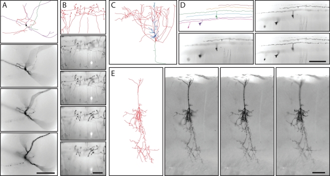
Figure 4. Tracing the morphologies of diverse AP expressing neurons from thick coronal sections of adult brains from various IRES-CreER;Z/AP mice.
A, Multipolar neuron in the inferior cerebral cortex. B, Mossy fibers in the cerebellum. C, The cortical pyramidal cell shown in Figure 3B. D, Cerebellar granule cells. E, a compact cortical pyramidal cell. A–D are from NFL-IRES-CreER;Z/AP brains, and E is from a ChAT-IRES-CreER;Z/AP brain; the tracing of the cell in E is shown at lower magnification in Figure 5J. A, B, D, and E are traced from 300 um thick sections, and samples from the corresponding Z-series bright field images are shown. C is traced from a 200 um thick section; a sample from the Z-series image is shown in Figure 3B. Scale bars: 0.2 mm.

