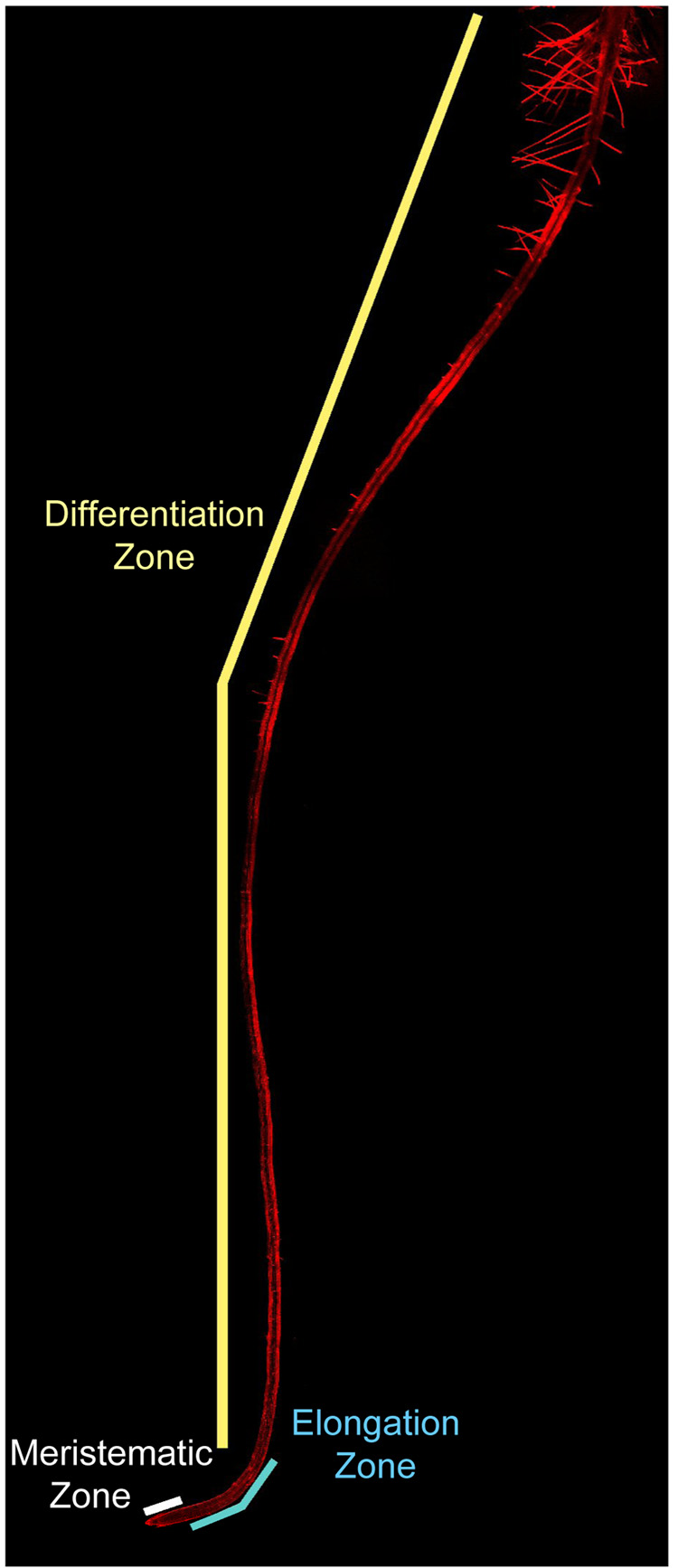
Figure 1.
The Arabidopsis primary root (6 days old) as viewed by laser confocal microscopy under 10X magnification. Three distinct zones mark the longitudinal axis of the root, namely, the meristematic (white bar), elongation (blue bar), and differentiation (yellow bar) zones. A close up of the meristematic and elongation zones is shown in Figure 3. Cell walls are visible in red from propidium iodide staining.

