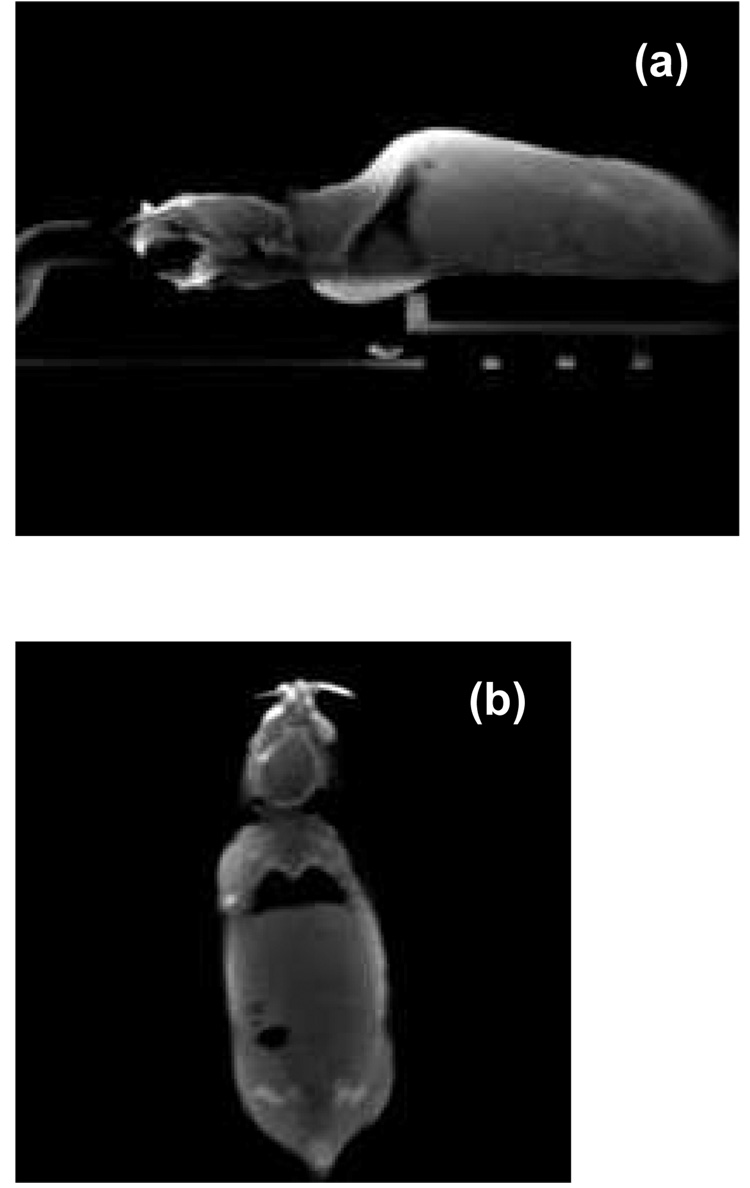
Figure 5.
(a) A sagittal slice of cone-beam CT of an anesthetized mouse scanned in the prone position. (b) A coronal CBCT slice of the same mouse. The mouse was immobilized with a custom head holder equipped with ear-pins and bite-block. The projection images were acquired with the flat panel detector.

