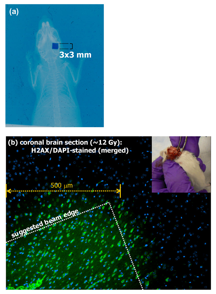
Figure 8.
(a) A double-exposed EBT film of the mouse irradiation where a single 3 mm × 3mm beam was directed posteriorily to the right hemisphere. (b) A merged image of the sectioned mouse brain that was stained with DAPI for cell nuclei and with antibody against γ-H2AX for correspondence with radiation-induced DNA strand breaks. The entire section shows DAPI staining, while there is an apparent sharp demarcation of a region that also shows γ-H2AX staining. A beam edge is suggested on the image as the experiment did not include geometric validation of the coincidence of the irradiation and γ-H2AX regions. The inset in the figure shows the extraction of the irradiated mouse brain for staining.

