
Figure 1. Degradation of overexpressed torsinA in PC6-3 inducible cell lines.
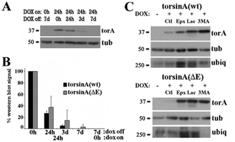
(A) Representative western blot of PC6-3 torsinA(wt) inducible cell lines that were induced with DOX for 24 h, DOX was then removed from the media and torsinA levels assessed by western blot at the indicated times. (B) Quantification of western blot signal normalized to loading control from three experiments as in (A) for torsinA(wt) and torsinA(ΔE). (C) PC6-3 cells were induced to express torsinA for 24h, DOX was removed from the media and treated as indicated for another 24h followed by western blot analysis. Increased ubiquitin immunoreactivity was only found after proteasomal inhibition. Ct: control; Epx: epoxomycin; 3MA: 3-methyladenine; Lac: lactacystin; torA: torsinA; tub: α-tubulin; ubiq: ubiquitin.