
Figure 2. Spheroid bodies are degraded through autophagy.
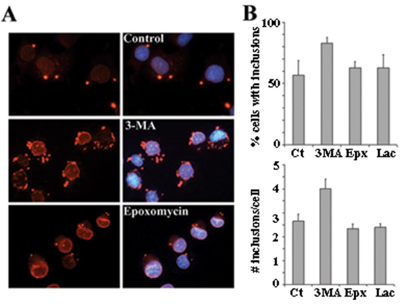
(A) IF analysis of PC6-3 cells inducibly expressing torsinA(ΔE) and treated as in figure 1C with the indicated drugs. TorsinA is seen in red and nuclear DAPI staining in blue. (B) Quantification of percentage of cells with spheroid bodies and average number of spheroid bodies per cell in randomly selected fields from three independent experiments under the indicated treatment conditions. Ct: control; Epx: epoxomycin; 3MA: 3-methyladenine; Lac: lactacystin.