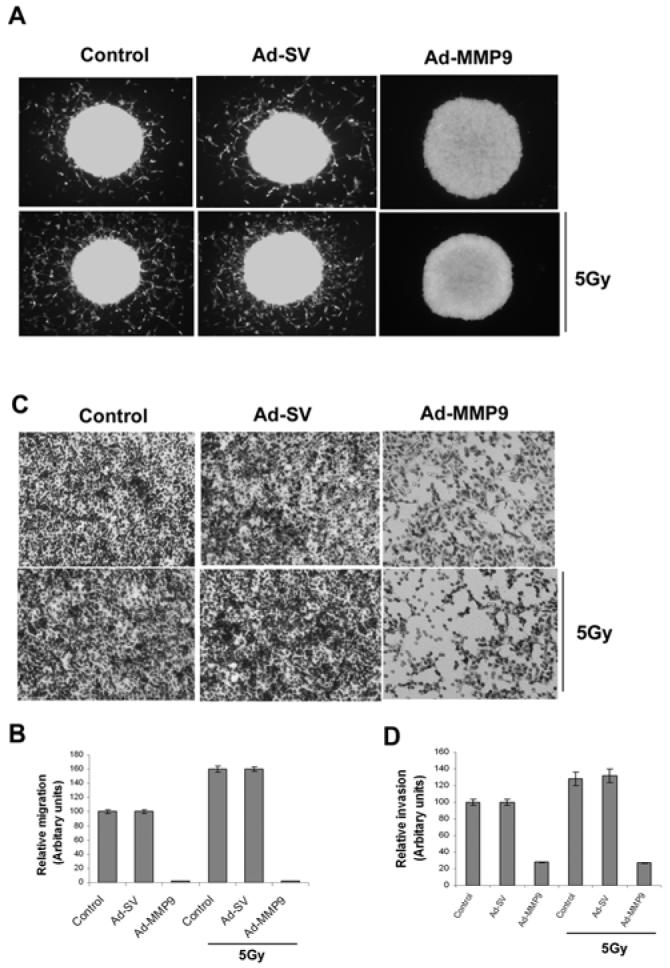
Figure 2. Knockdown of MMP-9 with Ad-MMP-9 reduces spheroid migration and invasion.

(A) Fluorescent-labeled IOMM-Lee cells were cultured in 96-well low attachment plates at a concentration of 7×10⁴ and spheroids were allowed to grow for 24 h at 37°C with shaking at 40-60 rpm. The spheroids were then infected with Ad-SV or Ad-MMP-9 and irradiated after 24 h. Untreated spheroids were also maintained to serve as the control (mock). Immediately after radiation treatment, the spheroids were transferred to 8-well chamber slides and maintained for another 24 h in serum-free media. Spheroid migration was analyzed by taking pictures using a fluorescent microscope. (B) Cell migration from the spheroids was quantified as the distance cells migrated from the spheroids. Values are mean ± SD from three different experiments (p<0.05). (C) IOMM-Lee cells were infected with Ad-SV or Ad-MMP-9 and irradiated at 5 Gy after 24 h of infection. Untreated (mock) were also maintained to serve as the control. After irradiation, the cells were trypsinized and 1×10⁵ cells from each treatment group and controls were cultured in the upper chamber of a transwell insert coated with matrigel (1 mg/mL). (D) We counted the number of cells in three different fields for each sample. The percentage invasion of cells treated with Ad-MMP-9 was analyzed and compared with the untreated (mock) cells. The graph represents the percentage invasion shown by cells infected with Ad-MMP-9 in comparison with untreated cells (mock). Values are mean ± SD from three different experiments (p<0.05).
