
Figure 5. MEK1/2 inhibitor and Dn-ERK constructs attenuate the phosphorylation of ERK and Akt.
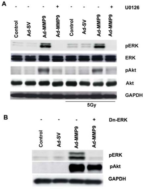
(A) IOMM-Lee cells were infected with 50 MOI of Ad-SV or Ad-MMP-9 and then treated with U0126 (20 μM). After 24 h of infection and treatment, one group of cells was irradiated at 5 Gy. After 16 h of irradiation, cell lysates were analyzed for total ERK, Akt and phospho ERK and Akt levels by Western blot analysis using the respective antibodies. GAPDH served as a loading control. (B) IOMM-Lee cells were transfected with 2ug/mL of Dn-ERK plasmid and infected with 50 MOI Ad-SV or Ad-MMP-9. Cell lysates were subjected to Western blotting with phospho-specific antibodies of ERK and Akt.