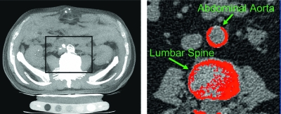
FIG. 1.
Abdominal CT (left) with analysis of vascular calcification (right) in the abdominal aorta. An Agatston score is determined by the product of the calcified lesion area (at least two contiguous pixels with a CT density at least 130 HU) and a co-factor dependent on the peak CT density (1, 130–200 HU; 2, 201–300 HU; 3, 301–400 HU; 4, >400 HU) of the lesion.

