Figure 1.
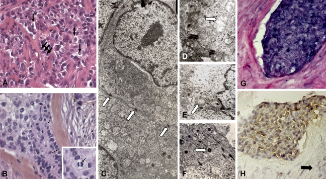
Structural and molecular results of the clear cell odontogenic carcinoma (CCOC). Hematoxylin–eosin–stained section of the clear cell odontogenic carcinoma shows the presence of epithelial nests with peripheral palisade cubical cells (A, black thick arrows) surrounding small compact interfaces. The presence of clear cells (B, black thin arrows) with atypia and mitosis, associated with hyalinized stroma containing giant cells, makes the diagnosis of CCOC more evident. Islands and strands of the clear cells are supported by a fibrous connective tissue stroma (B). Higher magnification of tumor cells shows a clear cytoplasm with uniform vesicular nuclei. (Inset: mitosis.) Desmosomes (C, arrows), endoplasmic reticulum, free ribosomes, glycogen rosettes, mitochondria (D, arrow), plasma membrane microvilli (E, arrow), and lysosomes (F, arrow) are ultrastructurally noted. Many cells exhibit a paucity of cytoplasmic organelles with prominent vacuolization. In the epithelial cells, amelogenin transcripts (G) and protein (H) are, respectively, detected by ISH and IHC. No amelogenin expression was detected in stromal cells (H, black arrow).

