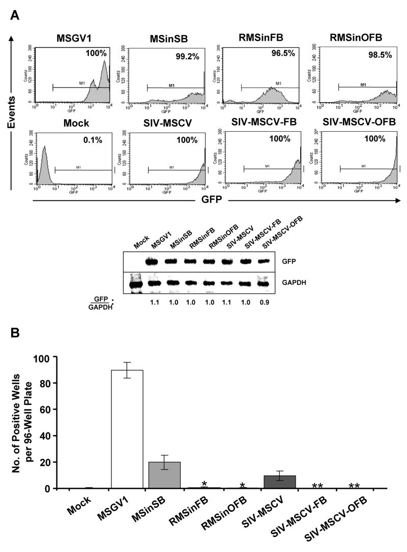
Figure 5.
Incorporation of FB element in SIN gammaretroviral and lentiviral vectors blocks in vitro transformation of murine BM cells. (A) Flow cytometric histograms of GFP fluorescence 1 week after the last transduction with the indicated vectors at an MOI of 10 (x 3). Shown in each case is the percentage of GFP-positive transduced BM cells. Below the histograms is a Southern blot analysis of NcoI/EcoRI (for gammaretroviral vectors) or NcoI/NotI (for lentiviral vectors)-digested genomic DNAs hybridized with a GFP probe (top panel) and a GAPDH probe (bottom panel) showing the relative vector copy number per cell population. The results presented are representative of two independent experiments. (B) Replating ability of vector-transduced BM cells. After retroviral transduction, BM cells were expanded as mass cultures for 2 weeks in IL-3-conditioned medium. During this period, cell density was adjusted to 1 x 10⁶ cells/ml every 3 days. Expanded BM cells were subsequently transferred into 96-well plates (n = 6 per vector) at a density of 100 cells/well. Two weeks later, the number of wells that contained proliferating cell populations were scored. Under these conditions, the majority of untransduced BM cells failed to survive replating or had ceased proliferating by this time. *Significantly fewer positive wells than MSinSB-transduced samples (P = 0.008). **Significantly fewer positive wells than SIV-MSCV-transduced samples (P < 0.05).

