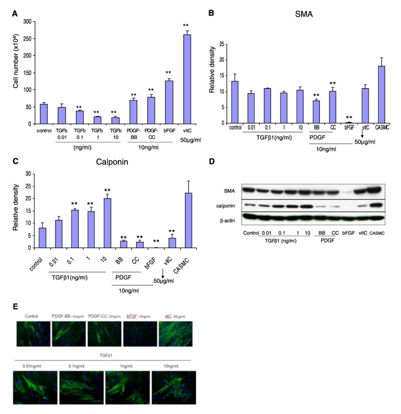
Figure 4.

Effect of various factors on hMSC cell proliferation and differentiation. hMSCs were seeded at 5.6 × 10³/cm2 in DMEM plus 5% FBS medium with one of the following supplements: 0 (control), 0.01, 0.1, 1, or 10 ng/ml TGFβ1; 10 ng/ml PDGF-BB, PDGF-CC, bFGF; or 50 μg/ml ascorbic acid. A) After 7 days, the cells were enumerated with 3% acetic acid with methylene blue on a hemacytometer (**P<0.05 vs. control; n=5). B, C) Western blots were performed on the protein lysates from each treatment group on SMA (B) and calponin (C; **P<0.05 compared to control, n=3). D) Representative Western blot on SMA, calponin, and β-actin. E) Immunofluorescence staining for SMA in hMSC culture after treatment with TGFβ1, PDGF-BB, PDGF-CC, bFGF, and vitamin C in addition to control for 7 days. Concentration of each factor is labeled in the figure.
