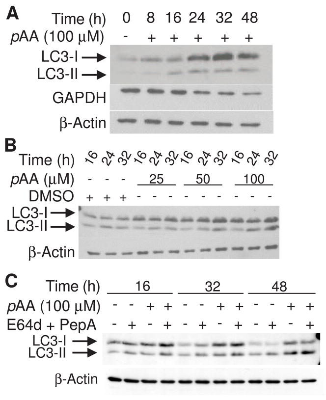
Fig. 7.
pAA-induced LC3 processing. MCF10A cultures were treated with nothing or 100 μM pAA (A) or DMSO and 25, 50 or 100 μM pAA (B) for varying times prior to being harvested and assayed by western blot analysis for expression of LC3-I and -II, GAPDH or β-actin. (C) Cultures were treated with nothing, 100 μM pAA, and/or 10 μM E64d plus 1 μM acetyl pepstatin A. Cultures were harvested 16, 32 and 48 h after pAA addition. E64d and acetyl pepstatin A were added 30 min prior to the addition of pAA. Each lane contained 40 μg of protein.

