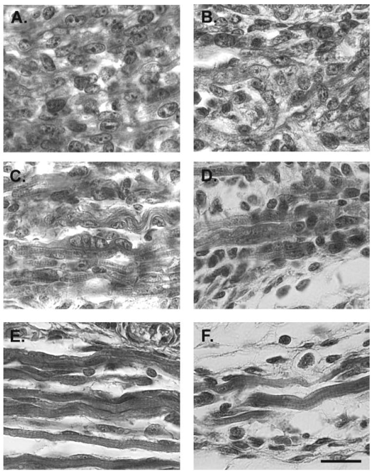
Figure 1.

Photomicrographs of trichrome-stained LA muscles in male (left) and female (right) mice on E16 (A, B), P1 (C, D), and P5 (E, F). (A, B) At E16, the LA in both sexes is composed primarily of myocytes and a few early myotubes. (C, D) Myotubes with centralized nuclei are seen at P1. Striations can also be seen at this age. (E, F) By P5, LA muscle fibers in both sexes have a mature appearance with peripheral nuclei and clear striations. Fibers are sparse in females, however, and some appear to be truncated. Nonetheless, those fibers that are present in females are similar in size and appearance to those in males. Scale bar = 20 μm.
