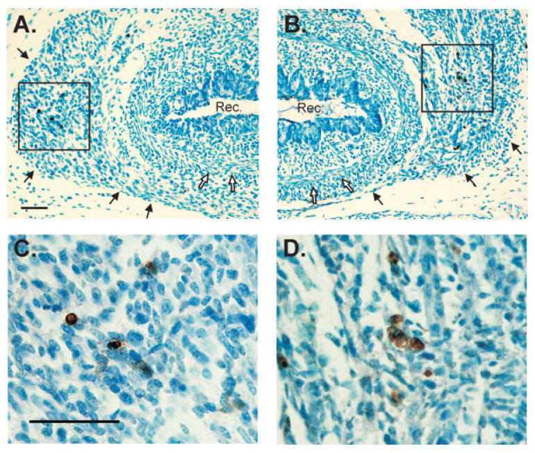
Figure 3.

(A, B) Low magnification photomicrographs of TUNEL-labeled sections through the perineum of an E18 male (A) and E18 female (B). Sections were counterstained with methyl green. Black arrows indicate the LA muscle; white arrows indicate smooth muscle of the rectum. Dorsal is down. (C, D) Higher magnifications of boxed areas in (A) and (B), respectively. A higher density of TUNEL-positive cells (brown) is seen in the LA of females. Scale bars = 50 μm. Rec, rectum. [Color figure can be viewed in the online issue, which is available at www.interscience.wiley.com.]
