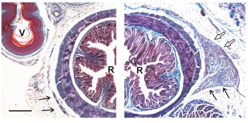
Figure 5.

Low-power photomicrographs of trichrome-stained sections through the perineum of an adult wild-type female (left), and a bax/bak DKO female (right). Black arrows point to the LA muscle, which is much larger in bax/bak DKO females than in wild type. White arrows (right) indicate a portion of the BC muscle in the bax/bak DKO. At this level of the perineum, the vagina (V) was present in wild-type females, but absent in bax/bak DKO females (see Lindsten et al., 2000). R, rectum. Scale bar = 300 μm. [Color figure can be viewed in the online issue, which is available at www.interscience.wiley.com.]
