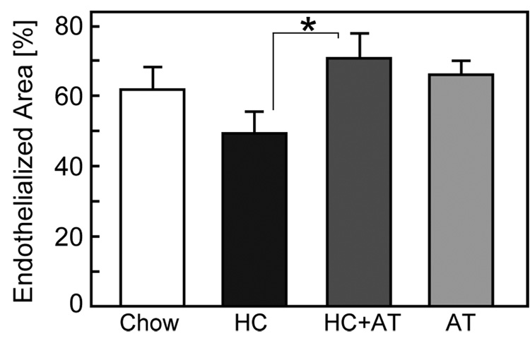
Fig. 2.
Endothelialization of ePTFE grafts. The endothelialized area and total luminal area of the graft were determined by SEM using the Scion Image analysis program. Endothelialization of ePTFE graft was reported as the percentage of vascular graft covered by endothelium relative to the total luminal area of the graft and expressed as the mean ± SE for each of the four dietary groups. (Chow: n=10 rabbits; HC: n=10 rabbits; HC+AT: n=5 rabbits; AT: n=5 rabbits) *P<.05.

