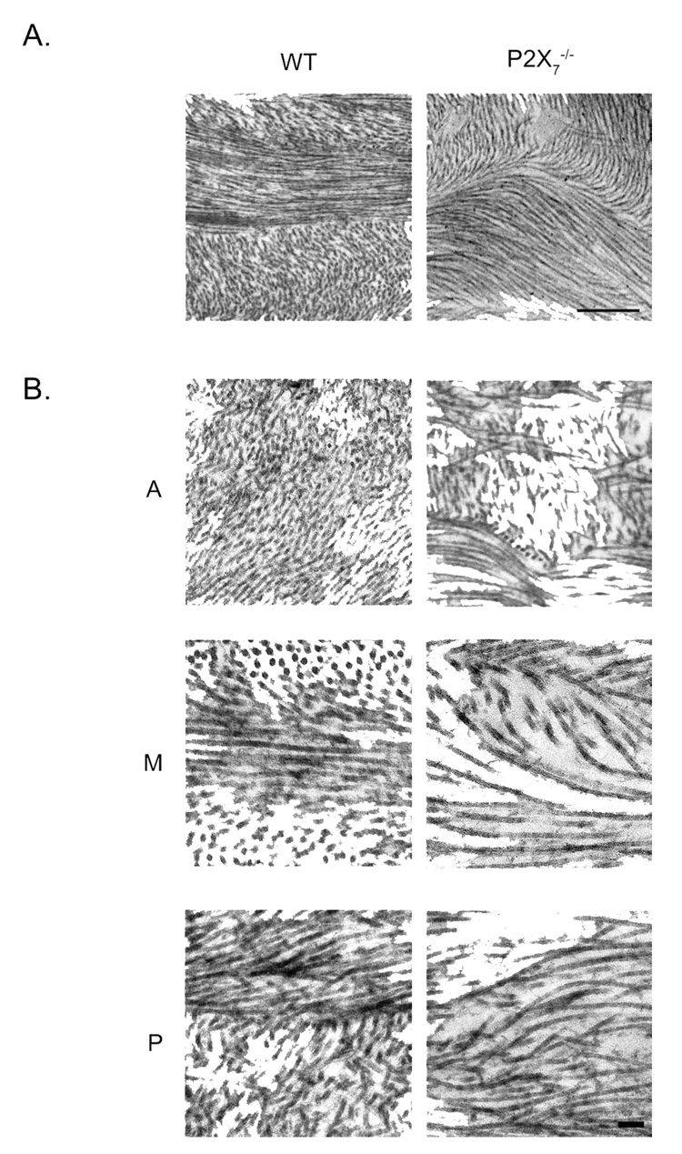
FIGURE 5.
Electron micrographs of central stromal collagen from unwounded WT and P2X7 −/− mice. (A) Low magnification of middle stroma depicts swirling of fibrils in P2X7 −/−. (B) Higher magnification of three distinct regions. A, anterior; M, middle; P, posterior. Scale bar: (A) 500 nm; (B) 100 nm.

