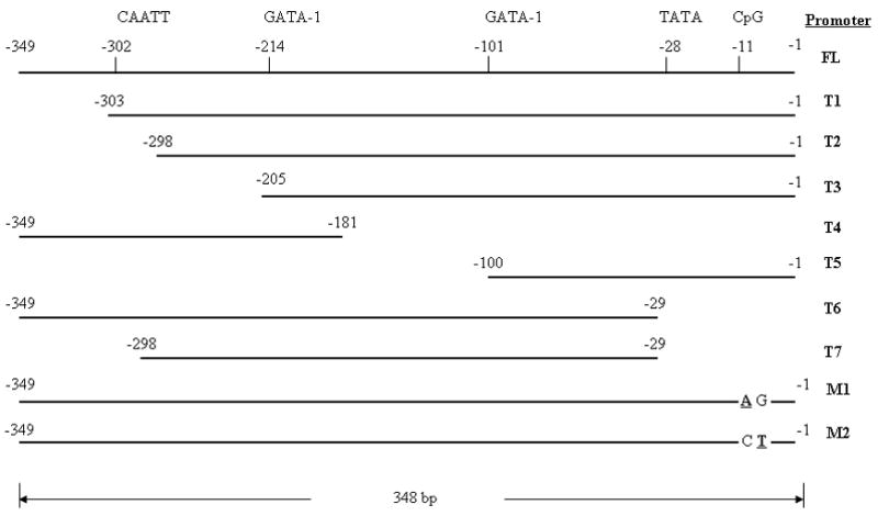
Figure 1.

A panel of truncated SEMG I promoters (T1, T2, T3, T4, T5, T6 and T7), including the positions of the putative GATA-1 and CAATT protein binding domains and the CpG dinucleotide, and a pair of mutated (in bold) full length SEMG I promoters (M1 and M2).
