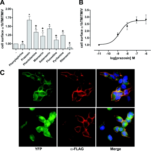
Figure 2. The α1-adrenoceptor antagonist prazosin promotes cell surface delivery of FLAG-α1b-adrenoceptor TMI-TMIV-eYFP.
(A) Intact cell anti-FLAG ELISA screening of the effect of several drugs known to have affinity for the α1b-adrenoceptor. ELISA assays were performed on cells stably expressing FLAG-α1b-adrenoceptor TMI-TMIV-eYFP after an overnight treatment with 10−5 M of each drug. Basal (−) staining reflects non-specific labelling of the cells. Statistics were performed using a one-way ANOVA with the application of the Tukey post-test analysis: *significantly greater than basal, +, significantly lower than the effect of prazosin, P<0.01 in each case. (B) Prazosin increases cell surface FLAG-α1b-adrenoceptor TMI-TMIV-eYFP in a concentration-dependent manner. Intact cell, anti-FLAG ELISA assays were performed as in (A) following overnight treatment with varying concentrations of prazosin. Results are expressed as mean of the fold increase of the absorbance detected in untreated cells (±S.E.M.). (C) Anti-FLAG immunocytochemistry (α-FLAG, red) was performed as in Figure 1 in fixed, non-permeabilized cells expressing either FLAG-α1b-adrenoceptor-eYFP (upper images) or FLAG-α1b-adrenoceptor TMI-TMIV-eYFP (lower images) that were treated overnight with 10−7 M prazosin. Imaging of eYFP (green) confirmed expression of both constructs and merging of the images (Merge) confirmed that prazosin treatment promoted cell surface delivery of FLAG-α1b-adrenoceptor TMI-TMIV-eYFP (lower panels, right-hand image). Blue staining represents nuclear staining with the DNA-binding dye Hoechst 33342.

