Figure 2.

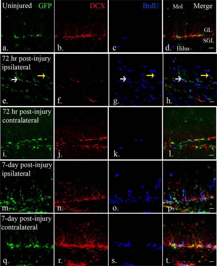
The distribution and response of neural progenitors in the adult dentate gyrus after CCI injury. a–d, eGFP- and DCX-expressing cells were found in the subgranular and granular layers of the dentate gyrus. BrdU-labeled cells were rarely seen in the dentate gyrus in uninjured mice. (e–h, After injury (72 h), eGFP-expressing early neural progenitors were detected in the subgranular layers (white arrow), as well as granular layers (yellow arrow). There is an apparent decrease of DCX-expressing late neural progenitors in the dentate gyrus in the ipsilateral hemispheres (f) at the same time that abundant BrdU-labeled cells were found in the dentate gyrus (g). (i–l) In the contralateral dentate gyrus, there is minimal apparent activation of GFP-expressing cells that mimics the control dentate gyrus. m–p, Robust numbers of eGFP-expressing cells persist in the ipsilateral dentate gyrus 7 d after injury (m). DCX-expressing late neural progenitors are more recovered at this time point (n) and there remains a large number of BrdU-positive cells (o). q–t, There is also an apparent increase in cell number and proliferation of both eGFP (q) and DCX-expressing (r) cells in the contralateral dentate gyrus 7 d after injury. Scale bars, 20 μm. Mol, Molecular layer; SGL, subgranular layer; GL, granular layer.
