
Figure 1.
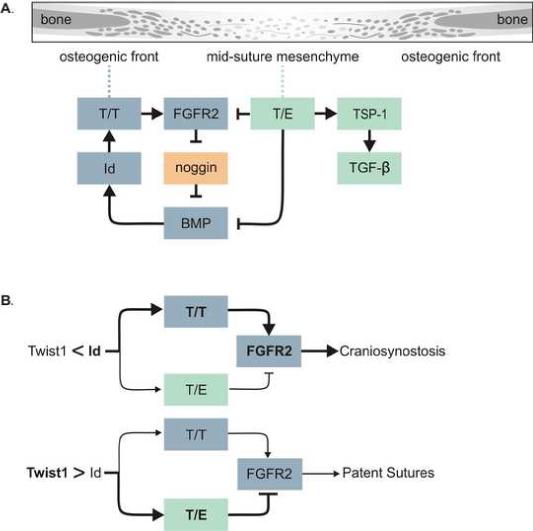
Model. (A) Drawing showing parts of a suture going from a more differentiated bone to a less differentiated mid-suture mesenchyme. The model shows T/T dimers up-regulating FGFR2 expression resulting in increased FGF signaling toward the middle of the suture. FGF signaling inhibits the expression of the BMP antagonist noggin, resulting in increased BMP signaling which up-regulates Id1 expression, further enhancing T/T homodimer formation. On the other hand, T/E dimers inhibit FGFR2 expression, as well as BMP signaling through binding to Smad proteins. T/E dimers also up-regulate TSP1 expression, which activates latent TGFβ. This could have differing outcomes dependent on which TGFβ isoform is present. (B) The ratio of T/T to T/E determines the functional output of Twist1 expression, and this is determined by the relative expression of Twist1 and Id1 proteins. When Id1 levels are greater that Twist1 this balance is shifted toward T/T formation resulting in up-regulation of FGFR2 leading to craniosynostosis. When Twist1 levels are higher than Id1 the balance is toward an increase in T/E formation, inhibiting FGFR2 expression and resulting in patent sutures.