
Figure 6.
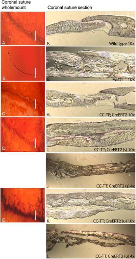
Expression of TT promotes overgrowth of the calvaria bones. (A-L) Mice were treated at P3 as in Figure 4 and were then harvested for analysis at 5 weeks of age. Skulls were stained with alcian blue and alizarin red and analyzed by wholemount (A-E) and sectioning (F-L). The number of mice treated was wild type (n=9), CC-TE (n=19), and CC-TT (n=8). Coronal sutures appeared patent in wild type mice (A), while Twist1+/− sutures were fused (B). TT expression resulted in varied degrees of two phenotypes: CC-TT (u) presented with an undulate phenotype (D), and CC-TT (s) had a serrate phenotype, which appeared to be tightly closed (E). The TE-expressing sutures were patent and showed greater separation between the bones (C). Cross-sections of coronal sutures confirmed the difference between the sutures: (F) coronal sutures from wild type adult mice showed a defined opening between the parietal and frontal bones; (G) Twist1+/− coronal sutures were clearly fused and the resulting bone was thicker than wild type; (H) CC-TE;CAG-CreERT2 sutures showed an increased mid-suture space with thinner bone ends compared to wild type; (I-L) CC-TT;CAG-CreERT2 expressing sutures of both serrate (s) (I,J) and undulate (u) (K,L) phenotypes were significantly expanded with severely overgrown calvaria bones that resembled the thickness of the Twist1+/− calvaria. These sutures are also shown using 4× magnification objectives to be able to view the entire sutures (J,L).